Percutaneous biopsy under ultrasound guidance is now well accepted and extensively used with a very low complication rate [1]. However there are very few reports in the literature of its application in the diagnosis of cardiac pathology. We report a case of percutaneous transatrial fine needle aspiration (FNA) o f a right a t r i a l tumour using a subxiphisternal approach.
CASE REPORT
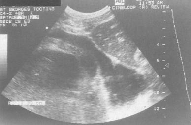
A previously healthy 24-year-old man presented to his General Practitioner with a short history of a dry cough and shortness of breath on exertion. Subsequent chest-radiography showed a markedly enlarged, globular cardiac silhouette, and transthoracic echocardiography showed a massive pericardial effusion and a large right atrial mass. A pericardial drain was inserted yielding over four litres of heavily blood-sustained fluid. Cytological examination revealed that the fluid contained abundant lymphocytes although immuno-phenotypic markers indicated that these were a normal inflammatory population of B- and T-cells with no evidence of clonatity. No malignant cells were seen and no microbial growth was obtained from the fluid after prolonged culture. Further investigation with transoesophageal echocardiography, computed tomography (CT) and magnetic resonance imaging (MRI) demonstrated that the mass infiltrated the free wall of the right atrium and extended around the aortic root and pulmonary trunk. There was no obstruction of either the superior or inferior vena cava (Fig. 1). MRI scan showed additional evidence of possible metastatic deposits in four vertebral bodies, although these were not apparent on either CT or isotope bone scan. The appearances of the mass were consistent with an angiosarcoma but histological confirmation was required. Transvenous biopsy was performed using a catheter bioptome (Cordis biopsy forceps) following localization of the tumour angiographically using contrast injected into the right atrium. Unfortunately the tissue obtained proved nondiagnostic despite taking multiple biopsies on two separate occasions and approaching the mass using the right femoral and right subclavian venous routes. Previous experience in our own institution suggests that open surgical biopsy of these highly vascular tumours is hazardous, and it was therefore decided to perform a percutaneous FNA under ultrasound guidance. Using an aseptic technique the right atrium was imaged in an oblique plane from the subxiphisternal region and a route plotted for the needle (Fig. 2). Under ultrasound guidance a 21 gauge
needle was directed into the lesion through the right atrial wall and the atrial cavity and aspirates were obtained. Three passes were made with the patient's consent. The specimens were immediately smeared, stained and examined under the microscope to determine the adequacy of the sample. Adequate material permitting more detailed cytological diagnosis was obtained from all three aspirates. Following appropriate stains, a diagnosis of angiosarcoma was confirmed. The procedure was welt tolerated with no immediate or late complications.

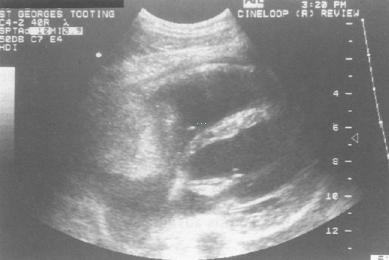
Fig. 1 Ultrasound image showing an echogenic mass lying within the right atrium.

Fig. 2 - The dotted line was used to assess depth of biopsy and to plan
the course of the needle through the right atrial wall, the atrial cavity
and into the mass. A picture of the needle in the mass was unfortunately
not obtained.
DISCUSSION
Primary cardiac angiosarcomas are the commonest form of primary malignant cardiac tumour making up
31% of the reported cases [2]. Patients with this tumour often present late with non-specific symptoms and over half the reported cases have metastatic spread at presentation [3]. The outlook for these patients is uniformly poor with a mean survival of between 6 and 9 months from the onset of symptoms [3-5]. In our patient, local excision of the turnout would not have been possible in view of its extension around the origin of the great vessels and MRI suggested metastatic spread to the spine. We were therefore keen to obtain a tissue diagnosis without recourse to open biopsy which has in our experience a considerable risk of complications. Pericardiocentesis proved unhelpful and other authors have reported this to be the case in the majority of patients [6]. Other reports have described successful
transvenous biopsy and its advantages [7, 8] but on this occasion we were unable to obtain diagnostic tissue using this technique.
Ultrasound guidance can be used to perform percutaneous biopsies and drainages of a wide variety of organs. It has several advantages over CT. No ionising radiation is involved and it can provide real-time images with imaging in multiple planes. This would be especially useful when scanning a dynamic structure such as the heart. Transoesophageal echocardiography has been successfully used to guide the biopsy of a right atrial lesion via the transvenous route [9] and percutaneous imaging has also been used to guide endomyocardial biopsies [10]. As far as we are aware this is the first description of a percutaneous transatrial approach for the biopsy of a right atrial mass. In addition, there is only
one case report of a cytological diagnosis of an angiosarcoma following FNA of a liver metastasis
[11]. In view of the anticipated difficulties in diagnosis, and since the patient did not experience any significant discomfort, three passes were made to increase the diagnostic yield and to permit multiple staining techniques to be carried out.
This is the first reported case of a percutaneous transatrial biopsy of a right atrial mass using ultrasound guidance. Three separate right atrial punctures were made on this occasion. The patient did not experience significant discomfort and there were no associated complications. It would appear that this technique may be a safer alternative to open cardiac biopsy.
REFERENCES
1 Matalon TAS, Silver B. US guidance of interventional procedures. Radiology 1990;174:43 47.
2 Loftier H, Grille W. Classification of malignant cardiac tumours with respect to ontological treatment (Review). Thoracic and Cardiovascular Surgeon 1990;38S:2:173-175.
3 Herrmann MA, Shankerman RA, Edwards WD et al. Primary cardiac angiosarcoma: A clinicopathologic study of six cases. Journal of Thoracic and Cardiovascular Surgery 1992;103:655-663.
4 Bear PA, Moodie DS. Malignant primary cardiac tumours: the Cleveland Clinic experience, 1956 to 1986. Chest 1987;92:860-862.
5 Dein JR, Frist WH, Stinson EB et al. Primary cardiac neoplasms: early and late results of surgical treatment in 42 patients. Journal of Thoracic and Cardiovascular Surgery 1987;93:502 511.
6 Glancy DL, Morales JB, Roberts WC. Angiosarcoma of the heart. American Journal of Cardiology 1968;21:413-419.
7 Adachi K, Tanaka H, Toshima H et al. Right atrial angiosarcoma diagnosed by cardiac biopsy. American Heart Journal 1988;115:482-485.
8 Hausheer FH, Josephson RA, Grochow LB et al. lntracardiac sarcoma diagnosed by left ventricular endomyocardial biopsy. Chest 1987;92:177-179.
9 Scott PJ, Ettles DF, Rees MR et al. The use of combined transoesophageal echocardiography and fluoroscopy in the biopsy of a right atrial mass. British Journal of Radiology 1990;63:222-224.
10 Di Pennestri F, Loperfido F, Salvatori MP et al. L'eco 2D nella guida a
 典型病例分享