1. Case report
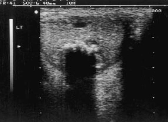
A 63-year-old man presented with a painless lump in his left testis that had first been noticed 4 months previously. A large mass (3 cm in size) was palpable in the lower pole of the left testis.There was no lymphadenopathy, palpable intraabdominal masses, nor gynaecomastia. Ultrasonography of the lesion emonstrated a hypoechoic solid mass posteriorly in the lower pole of the left testis containing multiple calcific foci associated with dense distal acoustic shadowing (Fig. 1).

Fig. 1. The appearance of the calcified epidermoid cyst on ultrasonography. A hypoechoic solid lesion is identified within the lefttestis (longitudinal section) that contains prominent calcified foci with ssociated distal acoustic shadowing.
No significant colour flow was identified within the lesion on colour Doppler ultrasound examination. Orchidectomy was performed and histology demonstrated a benign epidermoid cyst containing keratinising squamous epithelium and keratin debris with dystrophic calcification. There was no evidence of testicular intraepithelial neoplasia in the adjoining tissue.
2. Discussion
Epidermoid cysts in the testis account for approximately 1% of all testicular masses. They are benign, intratesticular lesions that contain keratinised debris or amorphous material and have a wall of fibrous tissue with an inner lining ofsquamous epithelium. They do not contain teratomatous elements in either the cyst wall or adjacent parenchyma (Price, 1969). Epidermoidcysts may represent a benign monodermal form of teratoma but no carcinoma in situ is found in adjacent seminiferous tubules, unlike typical teratoma (Coakley et al., 1998). They are often seen on ultrasound as well circumscribed, hypoechoic,encapsulated masses and calcification within them is rare (Reinberg et al., 1990; Eisenmenger et al.,1993). Although ultrasound is not diagnostic, when potential epidermoid cysts are identified, organ-preserving surgery can be planned. Exposing the testis via an inguinal approach, confirming the diagnosis of the lesion and the absence of in situ carcinoma in the adjacent tissue with frozen section histology may allow simple excision of the cyst instead of radical orchidectomy (Dieckmann and Loy, 1994). We have been able to find only one other reported case of an epidermoid cyst with similar calcified appearance on ultrasonography in the literature. Interestingly, the finding was made in a man of 52 years (Meiches and Nurenberg, 1991). Characteristically,epidermoid cysts are found in men between the ages of 20 and 40 years (Malek et al., 1986). Calcification may represent part of the natural history of a lesion that develops in earlier life.
 典型病例分享